

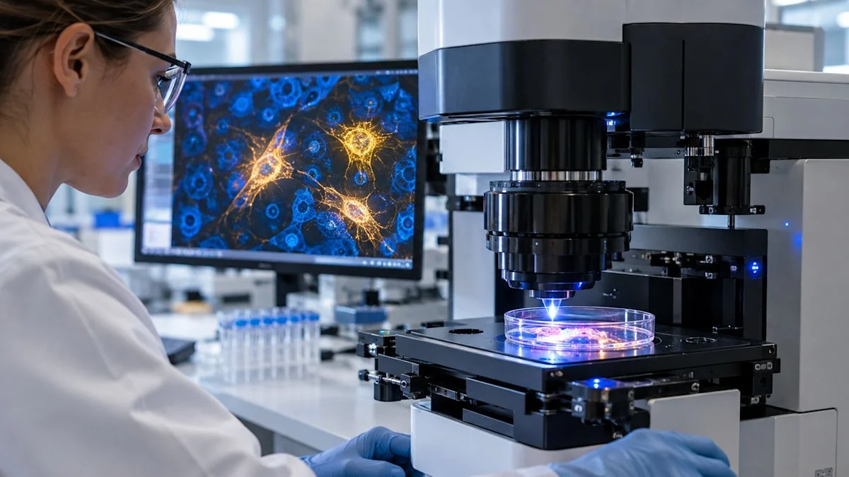
MIT researchers combined Raman microscopy and gene-expression data to identify signatures linked to senescent cells in mouse lung and skin. A machine-learning classifier turned the signals into a barcode; the method remains preclinical and unvalidated in people.
The samples came from 2- and 26-month-old mice. In both tissues, p21-positive senescent cells showed elevated Raman peaks at 1,131–1,135 inverse centimeters, linked to lipid-associated structures. Lung and skin also displayed distinct age-related gene programs.
The imaging system is not ready for routine medical use: analyzing about one square millimeter currently takes around 30 hours. Researchers are developing faster instruments focused on the most informative wavelengths; human tissues would also require extensive validation because senescent-cell states vary by organ, age and disease.
